Free 24/7 Nurse Advice Line: 844-GET-CHOC.
Fluoroscopy
A fluoroscopy exam is an imaging technique that uses continuous x-rays to create live images, almost like a movie, of anatomy and its function. While fluoroscopy might not be as familiar as a typical x-ray, we do these assessments daily to help diagnose abnormalities. They are often performed to assist in guiding invasive procedures, as well.
The fluoroscopic exams commonly performed at CHOC are:
- UGI (Upper Gastrointestinal Series): the X-ray camera records images of the esophagus, stomach, and the beginning of the small intestine (called the duodenum) while a child drinks a thick liquid (barium sulfate). Barium looks white on the images, and when it fills the organs of the GI tract, it makes them visible. Gas in the stomach and intestines looks black. The barium sulfate liquid looks like a light-colored milkshake and is often flavored for toddlers and young kids. It can be offered in a bottle or a cup with a straw. Occasionally, a child can’t drink the liquid, so it’s given through a small plastic tube or syringe.
- Small bowel follow through is used to see the entire small bowel. This is done by taking images of the patient’s abdomen every 30 to 60 minutes until the contrast reaches the large bowel. A small bowel follow through can last up to 7 hours. Please plan for this.
- Esophagram: this exam entails imaging of the esophagus and assessment of its function and anatomy. The patient will be given contrast orally (using a straw, bottle, or syringe). This is often done for esophageal narrowing, fistulas (TEF), difficulty swallowing, perforations (hole), obstructions etc.
- VCUG (Voiding Cystourethrogram): this exam is used to check the bladder, ureters, and urethra. The patient will have a catheter inserted by a registered nurse. We will use the catheter to fill the bladder with contrast. X-rays are then taken of the urinary tract. Once the bladder is full, the patient will be encouraged to urinate for the final images to assess the emptying of the bladder. This exam is often performed for urinary tract concerns including frequent urinary tract infections (UTI’s), Neurogenic bladder, bladder incontinence, vesicoureteral reflux etc.
- Contrast Enema: this exam is used to determine the function of the large bowel. A small tube (enema tip) is inserted into the rectum to allow contrast to move through the colon. X-rays are then taken of the intestines. This offers a clear view of the intestinal anatomy. This test may be done for chronic constipation, rectal incontinence, intussusception, and Hirschsprung’s disease.
- Fluoroscopy of the Chest (tube check): this exam is done to check for problems with a port or broviac. A registered nurse will access the lumen and inject contrast as the radiologist takes images. This is often done to check the position and length of the tube, and for lines with no blood return.
- Arthrogram: this exam is used to get images of joints (i.e. shoulder, elbows, ankles etc.). The radiologist will use medication to numb the area. Next they will use the x-ray guidance to slowly inject contrast into the joint. The patient will then get an MRI of the joint. This test checks the cartilage, ligaments and/or capsule of the joint.
- Lumbar Puncture: The radiologist will use medication to numb the area. With x-ray guidance the radiologist will use a needle to withdraw cerebral spinal fluid (CSF) for testing. Lumbar puncture is done to test performed CSF and for treatment of specific diseases.
- Myelogram: This test is done to see the bones and discs in the spinal column. In this exam, the radiologist will use medication to numb the area. With x-ray guidance, the radiologist will give contrast to help see the spine. Often, the patient will continue to CT for a scan of the anatomy. Performed but not limited to observing back pain due to injury vs. pathology.
- G/J- Tube placement check: This exam is done to check that a G/J tube is in the correct position and is working correctly. A technologist will give contrast through the tube and take images as the contrast enters the body.
- Modified Barium Swallow Study (MBSS): This study is performed with a feeding therapist. It is used to test how the patient is swallowing foods and liquids by adding barium to different foods and liquids. As the patient chews and swallow’s these foods and liquids, a radiologist or technologist will check the contrast passing from the mouth to the pharynx, and into the esophagus with special attention to the airway.
Can fluoroscopy harm my child?
A fluoroscopy exam is a safe, noninvasive procedure when done according to national safety guidelines. The benefits of fluoroscopy greatly outweigh the risk of harm when needed to diagnose illnesses and perform medical procedures. The radiology team at CHOC uses the least amount of radiation to obtain the necessary images.
What fluoroscopy safety guidelines does CHOC follow?
CHOC strives to minimize radiation exposure in children. We use techniques that result in shorter fluoroscopy times. We reduced radiation by up to 90 percent on fluoroscopic procedures.
What happens during fluoroscopy?

- Our radiology team will take you and your child to the exam area.
- The technologist will explain the procedure to you and your child and ask pre-screening questions.
- Your child will change into a comfortable gown for the procedure.
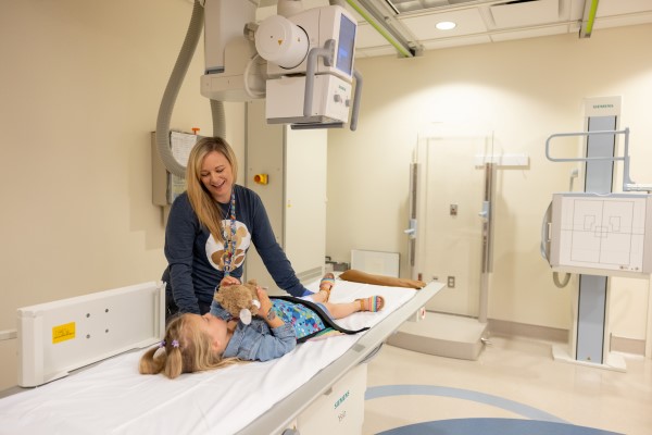
- Your child will lie on an exam table and a radiology technologist will position an X-ray machine (fluoro-tower) over the area being tested.
- The technologist will give the contrast to your child and will help your child move into different positions to get the images needed.
- Depending on the type of exam it usually takes 30 to 40 minutes to complete.
- It is important to note that this exam involves radiation, so only one caregiver will be allowed in the exam room with the child. Pregnant women are not allowed in the room during the exam. Siblings or anyone under the age of 18 are also not allowed in the exam room.
- It is also very important for your child to stay still during the exam. If your care team feels like your child will have difficulty with this, we will have our child life specialist close by to help with distraction
Will my child need a contrast material?
Fluoro exams require a contrast material. It helps the doctor get a better view of the studied body structure. Before your child’s appointment the radiology department staff will explain the procedure and the type of contrast being used.
Contrast may be given in a variety of ways depending on which area of the body the doctor is imaging. Some contrast taken by mouth enters the digestive tract. Other contrasts taken through a catheter enters the urinary tract. An enema is the contrast used to evaluate the rectum, or large intestine.
What types of Contrast are there?
At CHOC’s radiology department we use several different types of contrast. The contrast material used in our exams is based upon the exam type and pathology.
Water Soluble Contrast:
- OptiRay
- CystoConray / CystoGraffin dilute
- OmniPaque
Water soluble contrast us used for many of our studies. It is the safest measure for bowel, urinary tract, joint and spinal exams. In specific incidences it will be given orally.
Barium Contrast
Barium is the most used contrast for UGI’s and esophageal studies. It is a white material that is consumed in different consistencies. In certain studies, it is mixed with water and has a slightly sweet taste. In Multiple Barium Swallow Studies (MBSS), barium can be given in a dry, dust like form on different textured foods. It can also be given in a liquid form.
How should I prepare my child for fluoroscopy?
Helpful tips to help you prepare for your child’s fluoroscopy X-ray:
- Dress your child comfortably in clothes that are easily removed, such as sweats and a T-shirt.
- Bring special toys or books to help your child relax during the exam, or you may choose to use our toys.
- Avoid wearing jewelry and/or metal (zippers, snaps).
- Talk to your child honestly about the exam. Provide simple details about what will happen and they need to do.
Will my child feel any pain with fluoroscopy?
A fluoroscopy exam does not cause any pain. Sometimes your child may feel some discomfort with the method in which the of the contrast material is given. Our pediatric radiology team understand how to work with children. We will take all comfort measures possible, depending on the procedure. Someone from our radiology team will call you a day or two before the exam to explain the procedure in detail.
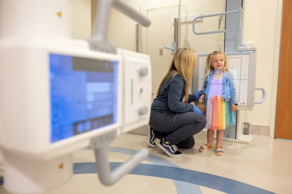
All our nurses, technologists and child life specialists use fun distractions like videos, toys, and activities to help children feel more at ease with the imaging process. Once patients are comfortable, our staff uses play medical equipment, pictures, and interactive tools to help children understand what to expect during the imaging exam.
What happens after the fluoroscopy?
- The nurse or technologist will give you any special instructions upon your departure.
- Intravenous or clear contrast (if given) will leave your child’s body through urine within 24 to 48 hours after the scan. The color of your child’s urine should stay normal.
- Barium, a thicker contrast, (if given), may produce white material in stool for two or three days. Barium can cause constipation (no stools or hard stools). Contact your child’s doctor if your child has not had a bowel movement after three days.
- After the exam, your child may eat or drink as usual, unless your child’s doctor tells you not to feed him.
How do I learn the results?
The radiologist will provide a report to the doctor who ordered your child’s fluoroscopy. The doctor will then discuss the results with you. Call your child’s doctor if you have any questions.
Why Choose CHOC?

- CHOC has been designated a Diagnostic Imaging Center of Excellence® (DICOE) by the American College of Radiology (ACR) for best-quality imaging practices and diagnostic care.
- CHOC uses only board-certified pediatric radiologists and specially trained pediatric radiology technologists, nurses and child life specialists.
- All radiology staff undergo age-specific training annually to learn how to work and communicate with children of varying ages.
- We are only one of a few medical centers in the country to have child life specialists working in a dedicated pediatric radiology and imaging department.
Meet Our Fluroscopy and X-Ray Director

Nahl, Daniel S. DO
Specialty:
Radiology
Appointments: 888-770-2462
Dr. Nahl is board certified by the American Board of Radiology with a certificate of added qualification in pediatric radiology.


